Achieving a beautiful, healthy smile is a journey, and X-rays are the roadmap that guides orthodontists along the way. These diagnostic images provide a detailed view of your teeth and surrounding structures, allowing orthodontists to monitor treatment progress and make informed decisions at every step.
But have you ever wondered how these seemingly simple images can provide such valuable insights? Let’s discover the power of X-rays in orthodontics – the tool that can help transform your smile into confidence at Hamer & Glassick Orthodontics.
The Role of X-Rays in Orthodontics
X-rays play a crucial role in orthodontic treatment by allowing orthodontists to:
- Assess the initial position of teeth and jaws
- Identify any underlying dental or skeletal issues
- Develop a personalized treatment plan
- Monitor the movement of teeth during treatment
- Detect potential complications early on
By capturing clear images of the teeth, roots, and supporting bone structures, X-rays enable orthodontists to make accurate diagnoses and tailor treatment plans to each patient’s unique needs.
Types of X-Rays Used in Orthodontics
Orthodontists typically use two main types of X-rays to track treatment progress:
1. Panoramic X-Rays
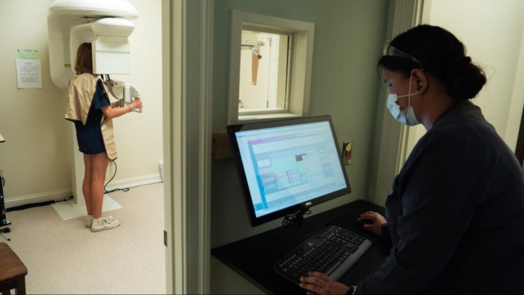
Panoramic X-rays, also known as Panorex or orthopantomograms (OPGs), provide a comprehensive view of your mouth, including all the teeth, jaws, and surrounding structures. This type of X-ray is taken using a specialized machine that rotates around the patient’s head, capturing a single, wide-angle image.
Panoramic X-rays are particularly useful for:
- Evaluating the position and development of wisdom teeth
- Identifying any missing or extra teeth
- Assessing the relationship between the upper and lower jaws
- Detecting any underlying pathology, such as cysts or tumors
2. Cephalometric X-Rays
Cephalometric X-rays, or Ceph X-rays, are side-view images of the head that show the relationship between the teeth, jaws, and facial structures. These X-rays are taken with the patient’s head positioned in a standardized manner, allowing for accurate measurements and comparisons throughout treatment.
Cephalometric X-rays are used to:
- Analyze the size and position of the jaws
- Determine the relationship between the teeth and facial profile
- Assess the growth pattern of the jaws and face
- Plan orthognathic surgery, if necessary

Tracking Progress with X-Rays
Orthodontists typically take X-rays at key stages of treatment to monitor progress and make any adjustments to your plan. These stages may include:
1. Initial Consultation
During the initial consultation, Dr. Hamer and Dr. Glassick will take a set of X-rays to assess the patient’s current dental and skeletal condition. These X-rays serve as a baseline for developing a personalized treatment plan and setting treatment goals.
2. Mid-Treatment Evaluation
As treatment progresses, orthodontists will take additional X-rays to evaluate the movement of teeth and the effectiveness of the chosen orthodontic appliances. These mid-treatment X-rays provide orthodontists the ability to make any necessary adjustments to the treatment plan, such as changing the type of braces or adding additional appliances.
3. Final Assessment
Once active treatment is complete, orthodontists will take a final set of X-rays to ensure that the teeth and jaws are in their desired positions. These X-rays also serve as a reference for future monitoring and help orthodontists determine the appropriate retention protocol to maintain the achieved results.
The Importance of Radiation Safety
While X-rays are an essential diagnostic tool in orthodontics, it’s important to minimize patients’ exposure to radiation. Modern X-ray technology, such as digital imaging systems, has significantly reduced the amount of radiation required to capture high-quality images. Orthodontists adhere to the ALARA principle, which stands for “As Low As Reasonably Achievable,” ensuring that the minimum possible radiation dose is used to acquire the required diagnostic details.
Orthodontists also use protective measures, such as lead aprons and thyroid collars, to shield patients from unnecessary radiation exposure during X-ray procedures. Patients can rest assured that the benefits of X-rays in monitoring orthodontic treatment progress far outweigh the minimal risks associated with the low levels of radiation used.

See Your Smile Transform
Your smile journey deserves precision at every step. With our advanced X-ray technology, you can witness the incredible transformation of your smile in vivid detail. From your initial consultation to your final, perfect smile, we’ll provide you with a comprehensive visual record of your orthodontic journey. Watch as your teeth gradually shift into their ideal positions.
Contact us now to start with a free consultation with Dr. Hamer and Dr. Glassick.
